A tear is found. Fibrin is placed.
The FibraDisc Procedure addresses a specific finding — a torn disc — by placing fibrin sealant into it through a needle. This page walks the whole thing, from the MRI you already own to the months after.
Evidence & regulatory status
Fibrin sealant is FDA-approved for surgical use. Its use inside the disc is an off-label use, which your physician will discuss with you during informed consent. The published evidence for intradiscal fibrin is pilot and observational data; one randomized trial did not meet its primary endpoint. Outcomes vary and are not guaranteed.
The outer ring tears. The centre leaks.
Most back and neck pain that comes from a disc is not about the disc's shape. It is about a tear in its outer ring and what seeps out through it. Three things to know before anything on this site makes sense.
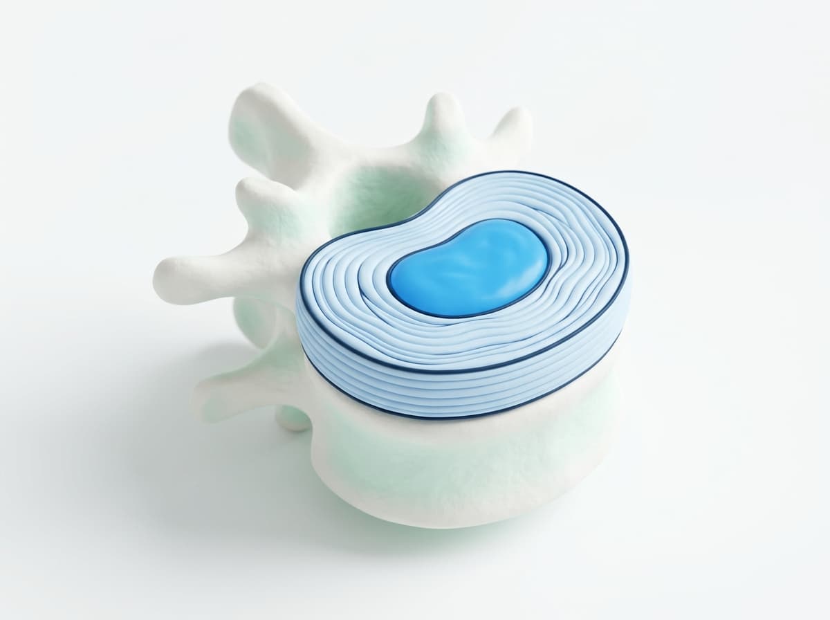
A cushion with a tough outer ring
Each spinal disc is a soft, gel-like centre held inside a ring of many thin, tough layers. The ring keeps the centre in place and carries the load every time you bend, sit, or lift.
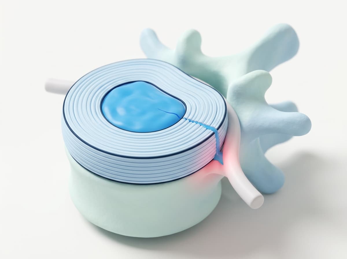
The ring can split, and the centre can leak
Wear, strain, or injury can tear the outer ring. Material from the centre seeps through the tear and can irritate the nerve root beside it — which is why a torn disc can hurt even when nothing is pressing on a nerve.
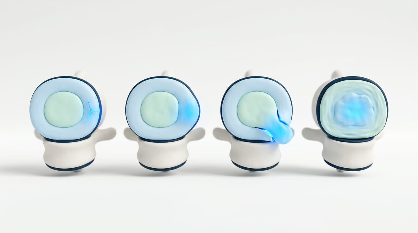
Often one problem, at different stages
A tear, a bulge, a herniation, a disc that has lost height — these are frequently the same disc at different points, not four separate conditions. That is why the useful question is not the label on your report, but whether a disc is torn and leaking.
Find the tear. Place the fibrin.
The tough outer ring of a spinal disc can split. Those tears leak, they hurt, and they do not reliably show up on an MRI. This procedure finds them with live imaging and places fibrin sealant into them — fibrin is the same clotting protein your body uses to close a wound.
Your MRI is reviewed
A physician reads the study you already have and tells you whether you look like a candidate — before you book anything.
An MRI is the entry requirement, not a formality. Without one, we can talk about the procedure in general terms but we cannot tell you anything about your spine. If you do not have a recent study, your primary care physician can order one.
An annulogram finds the tears
Contrast dye is introduced into each disc under live imaging. Torn discs leak. Intact ones do not.
This is the part an MRI cannot do. An MRI shows disc shape and height; it does not reliably show where the annulus has split. The annulogram shows the leak in real time, which is what determines which levels are treated — and sometimes shows that the disc everyone blamed is not the one that is torn.
Fibrin sealant is placed into the tears
Fibrin — a biologic sealant already used in surgery — is placed into the torn discs through a needle.
No incision, no hardware, no bone removed, and no disc taken out. Fibrin is the body's own clotting protein; in the disc it acts as a sealant and a scaffold. Because nothing structural is altered, the levels above and below are not loaded the way they are after a fusion.
You heal over months, not days
Most people are up and about the next day. The procedure is intended to support gradual healing, and the honest answer is that it takes time.
This is not a procedure that trades a long recovery for an instant result. You walk out the same day and resume ordinary activity quickly, but any healing that follows is measured in months, varies between individuals, and is not guaranteed. Anyone who promises you a switch being flipped is selling you something.
The MRI shows the shape. The annulogram shows the leak.
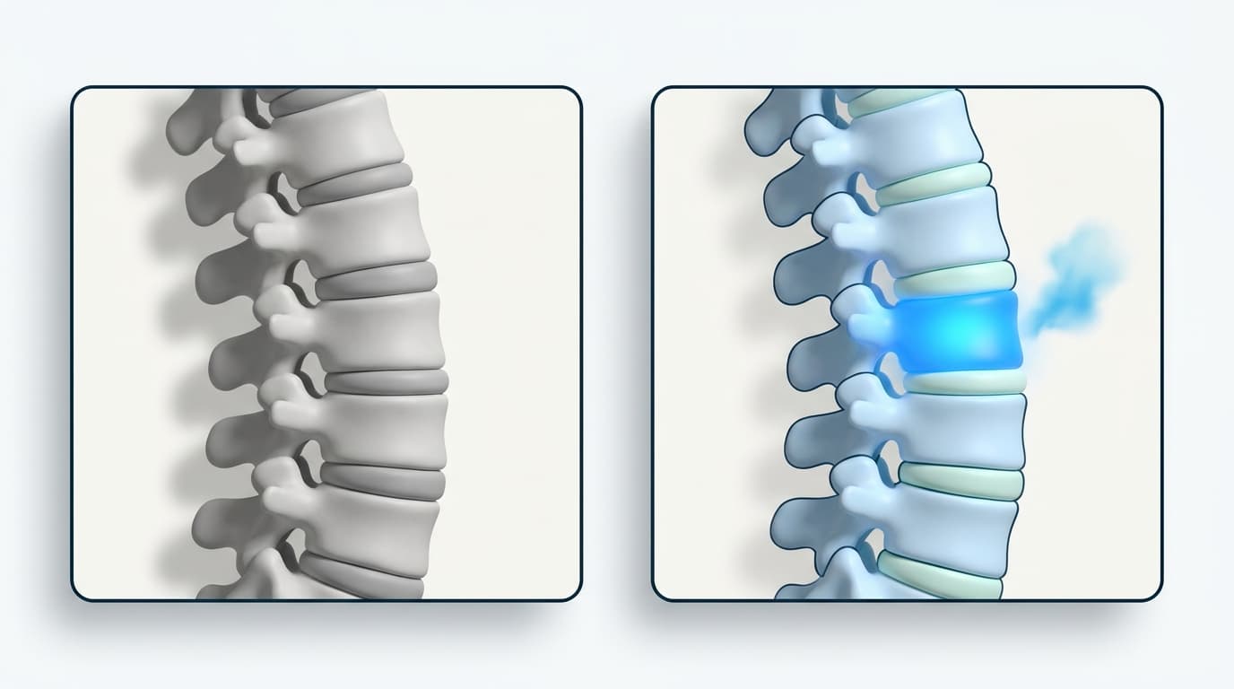
- MRI
- Disc shape and height. A tear that leaks often looks unremarkable here — which is why so many people arrive with a report that says nothing much.
- Annulogram
- Contrast introduced into each disc under live imaging. Torn discs leak; intact ones do not. This is what decides which levels are treated.
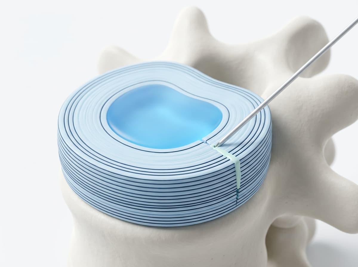
Then, the fibrin
Fibrin sealant is placed into the tear itself through a needle — no incision, no hardware, nothing removed. Same visit as the annulogram.
Illustrations, not patient imaging. Your own scan is read by a physician, in writing, before you travel.
What each option actually does to your spine.
No outcome claims in this table — only what is removed, what is left behind, and what remains possible afterwards.
Incision
- FibraDisc
- None — needle only
- Fusion
- Open or minimally invasive incision
- Artificial disc
- Open incision
Hardware left in the body
- FibraDisc
- None
- Fusion
- Screws, rods, cages
- Artificial disc
- Prosthetic disc
Bone removed
- FibraDisc
- None
- Fusion
- Yes
- Artificial disc
- Yes
Your own disc
- FibraDisc
- Kept
- Fusion
- Removed and fused
- Artificial disc
- Removed and replaced
Adjacent levels
- FibraDisc
- Not surgically loaded
- Fusion
- Load transferred to neighbouring discs
- Artificial disc
- Motion preserved at the treated level
Surgery still possible afterwards
- FibraDisc
- Yes
- Fusion
- Revision only
- Artificial disc
- Revision only
Setting
- FibraDisc
- Outpatient, same day
- Fusion
- Hospital, often overnight
- Artificial disc
- Hospital, often overnight
Typically covered by insurance
- FibraDisc
- No — self-pay
- Fusion
- Usually
- Artificial disc
- Usually
It is not right for everyone. We will say so.
Often a good fit
- Chronic low back or neck pain that has not resolved with conservative care
- A recent MRI showing disc degeneration, bulging, or herniation
- Pain after a failed spine surgery, including fusion
- You have been told fusion is the next step and want another option first
- You want to avoid hardware and preserve your own disc
Not a fit, or not yet
- No recent MRI — we cannot assess you without imaging
- Progressive weakness, foot drop, or loss of bladder or bowel control (seek care now)
- Active spinal infection or cancer involving the spine
- Severe spinal instability or high-grade slippage
- Expecting insurance to cover it — the procedure is self-pay
The things people ask before they call.
It is a non-surgical treatment for torn spinal discs. Under live imaging, a physician first performs an annulogram to find which discs are actually leaking, then places fibrin — a biologic sealant already used in surgery — into those discs through a needle. There is no incision, no hardware, and no bone removed.
Start with the scan you already have.
A physician screens the MRI you already have and tells you in writing whether you appear to be a candidate — before you spend anything, least of all a flight.